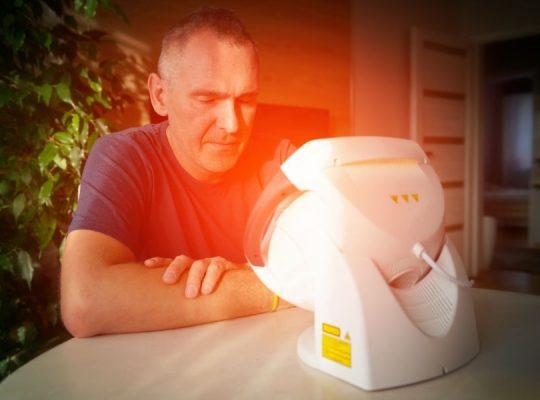
References to show Neuroimaging is a diagnostic for ME/CFS:
Neurological Dysfunction in Chronic Fatigue Syndrome, Journal of Chronic
Fatigue Syndrome (The Haworth Medical Press, an imprint of The Haworth
Press, Inc.) Vol. 6, No. 3/4, 2000, pp. 51-68.
Abhijit Chaudhuri, DM, MD,
MRCP; Peter 0. Behan, DSc, MD, FACP, FRCP
"SPECT Imaging of the Brain: Comparison of Findings in Patients with Chronic
Fatigue Syndrome, AIDS Dementia Complex, and Major Unipolar
Depression,"
Richard B. Schwartz, Anthony L. Komaroff, Basem M. Garada,
Marcy Gleit, Teresa H. Doolittle, David W. Bates, Russell G. Vasily,
B. Leonard Holman;
American Journal of Roentgenology, Vol 162, 943-951,
Copyright © 1994 by American Roentgen Ray Society.
Summary: "This study shows that CFS (ME) shares some similarities on
SPECT imaging with AIDS Dementia Complex acute changes in
radionuclide uptake in the younger population may be caused by inflammatory
processes at the cellular or micro vascular level .... the findings in
CFS (ME)
are consistent with the hypothesis that CFS (ME) ... results from a
viral infection
of neurons, glia or vasculature .....viral infection can provoke
neurological
dysfunction by interfering with intra-cellular mechanisms or membrane
transport
systems .... or by cerebral hypo perfusion due to vasculitis".
Gordon R et al. Cortical motor potential alterations in chronic fatigue
syndrome.
Int J Molec Med. 1999; 4: 493-99.
Proton magnetic resonance spectroscopy of basal ganglia in chronic
fatigue syndrome. Chaudhuri A, Condon BR, Gow JW, Brennan D,
Hadley DM. Neuroreport. 2003 Feb 10;14(2):225-8.
Costa DC, Brostoff J, Douli V, Eli PJ. Brainstem hypoperfusion in
patients with Myalgic Encephalomyelitis-Chronic Fatigue
Syndrome. Eur J Nucl Med 1992 19:733.
Brainstem perfusion is impaired in chronic fatigue syndrome. DC Costa,
C Tannock and J Brostoff. Quarterly Journal of Medicine December
1995:88:767-773)
Relationship of brain MRI abnormalities and physical functional
status in chronic fatigue syndrome Cook DB, Natelson BH.
Int J Neurosci 2000:107:(1-2):1-6
Brain positron emission tomography (PET) in chronic fatigue
syndrome: preliminary data Tirelli U et al. Am J Med 1998:105:
(3A):54S-58S
Brain MRI abnormalities exist in a subset of patients with
chronic fatigue syndrome. Lange G, DeLuca J, Maldjian JA,
Lee H, Tiersky LA, Natelson BH. J Neurol Sci.
1999 Dec 1;171(1):3-7.
Chronic fatigue syndrome--aetiological aspects. Dickinson CJ.
Eur J Clin Invest. 1997 Apr;27(4):257-67
Brain MR in chronic fatigue syndrome. Greco A, Tannock C,
Brostoff J, Costa DC. AJNR Am J Neuroradiol. 1997 Aug;18(7):
1265-9.
Relationship of brain MRI abnormalities and physical functional
status in chronic fatigue syndrome. Cook DB, Lange G, DeLuca J,
Natelson BH. Int J Neurosci. 2001 Mar;107(1-2):1-6.
Quantitative assessment of cerebral ventricular volumes in
chronic fatigue syndrome. Lange G, Holodny AI, DeLuca J,
Lee HJ, Yan XH, Steffener J, Natelson BH. Appl Neuropsychol.
2001;8(1):23-30.
Dr. Byron Hyde, a Canadian specialist in Myalgic Encephalomyelitis
offers this definition of Myalgic Encephalomyelitis: "Myalgic
Encephalomyelitis is a measurable, diffuse post-encephalitic illness.
The illness is characterized by (1) its acute onset,
(2) the diffuse, non-focal persisting nature of the encephalopathy,
and (3) the chronicity of the resulting symptoms. These symptoms
consist of the rapid exhaustion or loss of stamina of motor, sensory,
intellectual, and cognitive abilities. M.E. is of infectious/autoimmune
origin and less commonly, a toxic/autoimmune origin.
M.E. occurs in epidemics and sporadic cases."
[Note by Rich Van Konynenburg, Ph.D, who reported on Dr.
Hyde's lecture at the recent Wisconsin conference of medical practitioners
specializing in Myalgic Encephalomyelitis: "Basically, what he's saying
here is that Myalgic Encephalomyelitis starts with an inflammation of
the brain that occurs rather suddenly. This initial inflammation usually
results from an infectious/autoimmune process, but it can also be
caused by a toxic/autoimmune process. This sudden, short-term
inflammation is followed by a disorder of the brain that continues
over time. This chronic disorder of the brain is not localized to a
small part of the brain, but is spread out over large regions of the
brain, and it leads to chronic symptoms that can involve essentially
all the normal functions of the brain. Myalgic Encephalomyelitis
occurs both in epidemic-type clusters of cases as well as cases
that are occasional and isolated."] Dr. Hyde said that though the
primary injury in Myalgic Encephalomyelitis is the diffuse CNS
encephalopathy, the illness may cause or be associated with
measurable dysfunction in end organs and various body systems.
The most commonly injured end organs and systems are (1) the
thyroid gland, (2) the cardiovascular system and (3) the immune system.
The CNS dysfunctions are caused by widespread, measurable, diffuse
micro-vasculitis affecting normal cell operation and maintenance.
"The evidence would suggest that Myalgic Encephalomyelitis is
caused primarily by a diverse group of viral infections that have
neurotropic characteristics and that appear to exert their influence
primarily on the CNS arterial bed. The available brain technology
limits the viral site of action to the capillaries and microarterial CNS
bed. This diffuse vascular site of injury rather than a neurological
cellular site of injury explains the natural history of Myalgic
Encephalomyelitis-type illness." [Note--What he is saying here is that
there is evidence that the causes of Myalgic Encephalomyelitis are
any of a group of viruses that are able to infect the brain. By means of
high-resolution SPECT scanning, he can tell that they mainly affect the
small arteries and capillaries in the brain.] "It is also noted that many
Myalgic Encephalomyelitis patients also have generalized arterial
pathophysiology [Note --In other words, there are problems with
the arteries all over their bodies.], causing various vascular problems
that include in numerous patients: (1) insufficient blood pressure increase
on exertion, (2) hyperelasticity and hyper-contractibility of arterial blood
vessels, (3) various forms of arterial mediated vascular orthostatic
pathophysiology [Note --In other words, they have difficulty standing up
because of problems with their arteries] as demonstrated by Drs. David
Streeten, David Bell, and Peter Rowe, and (4) cholinesterase dysfunction
in the arterial wall, causing arterial elasticity dysfunction as
demonstrated
by Dr. Vance Spence at Dundee University, Scotland."
Brain Blood Flow
This article appeared in The Medical Post, January 19, 1993
Patients suffering from chronic fatigue syndrome (CFS) do so
because of cerebral function abnormalities as a result of diminution
of brain blood flow, says a California nuclear medicine specialist.
Dr. Ismael Mena of the Harbor-UCLA Center in Los Angeles said:
"All adult patients with chronic fatigue have abnormal diminution of
blood flow to different areas of the brain - mostly in the frontal lobes."
He told a meeting on CFS recently that this diminished blood flow
could reflect damage to those areas, but might also be the result of
damage to another area of the brain that is expressed in the frontal
lobes. He said the primary damage may be in the limbic system,
particularly in the hippocampus. "This is the one that projects
directly in the frontal lobes where most of the damage is seen in
patients with chronic fatigue."
The diminution of blood flow to the temporal lobes could also
explain why patients with chronic fatigue complain of cognitive
impairment as that is where the memory resides. Using brain
Single Photon Emission Computer Tomography (SPECT), Dr.
Mena studied the brain flow patterns of 19 normal people with
an average age of 66; 33 CFS patients with an average age of 55;
and 26 patients with depression and a mean age of 62.
Great Precision
Although computed tomography (CT) and magnetic resonance
imaging (MRI) can denote with great precision the structures of
the brain, with brain SPECT the functions of the brain can be
examined. Dr Mena said he examined the brain blood flow
because it is directly related to brain function. Two sets of
measurements were taken - one using 133Xe rCBF to measure
the amount of blood flow in the brain tissue, the other,
99mTc-HMPAO, to provide images of brain blood flow.
Dr Mena said in a normal person the blood flow, when
measured using 133Xe rCBF, fluctuates between 50 and
67 mL/min/100g of brain tissue. However, patients with CFS
don't exhibit a uniformity in blood flow. For example, in one patient,
aged 42, blood flow was 37 mL/min/100g in the right frontal lobe.
"For a person of this age it should be 55, ± 5", he said. In another
example, a 48 year-old woman, there was extensive hypoperfusion
in the frontal lobes. "But more extensively in the dorsal frontal lobes
with blood flow between 38 mL and 47 mL", he said. The
99mTc-HMPAO data also showed patients had both frontal
perfusion and temporal
hypoperfusion, but to provide a more precise analysis of the data,
Dr Mena said regions of interest were set and a computer
was used to calculate the amount of blood flowing in them.
Approximately 100 measurements were taken per patient.
Dr Mena said when this type of analysis was done, the distribution
of 133Xe rCBF in a normal elderly person is close to
45 mL/min/100g. Again, this data showed in patients with CFS
there was a diminution of blood flow in the right hemisphere. With
the 99m Tc-HMPAO data, Dr Mena said there was significant
hypoperfusion throughout the frontal lobes as well as the right
temporal lobe.
Dr Mena also examined the effect of exercise on patients with CFS.
Patients were given the standard cardiac stress test. While a normal
person usually gets his wind back within five minutes after doing the
test, those with CFS had to wait at least one hour before their levels
of carbon dioxide returned to their pre-exercise levels. "There's a
marked worsening of blood flow in both hemispheres, but mostly in
the right hemisphere, and these abnormalities persist up to 24 hours",
he said. After 24 hours, sometimes even 48 hours, patients' brain
blood flow returned to where it was at normal activity level.
Acknowledgment: Reprinted from The Messenger, March 1993,
newsletter of The M.E. Association of Canada.
Reprinted from Emerge, September 1993.
"America's Biggest Cover-Up by Neenyah Ostrom"
Chapter Twenty: CFS Patients Have A Brain Defect Similar To That Found In AIDS Dementia"
It has been known
for some time
that CFS patients have abnormal
blood flow in their brains; that is, some areas of the brain are not
getting as much blood as they should. Very recently, however, studies
of patients with AIDS dementia have shown that they, too, have
abnormal brain blood flow, raising the question of whether a similar
disease process is taking place in both sets of patients.
It has only been possible to measure blood flow in the brain with the
development, over the last few years, of very specialized types of brain
scan technologies. One type of brain scan that can detect blood flow
abnormalities is called SPECT (which stands for "single photon emission
computer tomography").
Dr. Ismael Mena has studied CFS patients' brains using SPECT scans
at the University of California-Los Angeles, where he is a professor of
radiology. Over several years' investigation, Dr. Mena has consistently
reported that 71 percent of CFS patients have a diminished flow of
blood in their brains.
Dr. Mena has also commented, at scientific conferences, that CFS
patients do not have equal blood flow on the two halves of their brains.
That is, when the blood flow on the right and left sides (or hemispheres)
of the brain were compared in individual CFS patients, the flow in the
two halves of the brain can differ by twice as much as they do in
healthy people.
This information suggests that some type of organic brain disease
may be a component of CFS. While it has long been recognized
that AIDS patients can develop very serious mental problems,
sometimes labeled "AIDS dementia," only recently have such
patients been studied using SPECT scans. The similarity between
"AIDS dementia" patients' SPECT scans and those of CFS
patients are striking.
SPECT scans in AIDS patients were discussed at the 1992
international AIDS meeting (in Amsterdam) by a research team
from the Houston Immunological Institute. Rather shockingly, they
found that exactly the same percentage of AIDS patients -- 7
1 percent -- as CFS patients had abnormalities in brain blood flow.
The Houston research team also found that more severe cases of
AIDS dementia showed greater abnormalities in brain blood flow,
and they suggested that SPECT scans could provide a very
important diagnostic tool for physicians treating patients with AIDS."
References assembled by Steven Du Pre
Website for National Alliance for Myalgic Encephalomyelitis:
http://www.name-us.org